A multi-modal analysis to characterise fibrotic tissues
Pulmonary fibrosis (PF) is a disabling disease characterised by progressive involution of the lungs, that currently has no available curative approaches. Most cases of PF are of the idiopathic type, a heterogeneous group comprising disorders with very different pathophysiology: it is then essential to improve tissue characterisation techniques so that each type of fibrosis can be addressed in the most appropriate way.
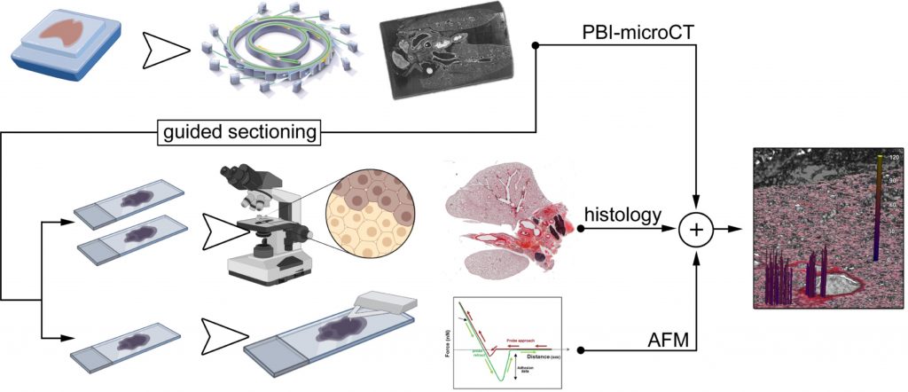
Our PhD fellow Lorenzo D’Amico, together with colleagues from University Medical Center Göttingen, Max Planck Institute for Multidisciplinary Sciences and Georg-August-Universität Göttingen Institute for X-Ray Physics, developed and applied a novel multi-technique characterization approach to tissues coming from two different preclinical mouse models, in which PF was induced either chemically (by Bleomycin) or by genetic modification. Tissue analysis was then based on the combination of three different techniques: classical histopathology, propagation-based phase-contrast micro computed tomography (PBI-microCT, available at the SYRMEP beamline of the CERIC Italian partner facility, in Elettra Sincrotrone), and atomic force microscopy (AFM).

Researchers were then able to discriminate samples from the Bleomycin-induced model from those of genetically altered mice, thus distinguishing the different pathophysiology leading to PF.
This new pipeline of investigation could represent a new approach to the analysis of tissue samples, since Formalin Fixed Paraffin Embedded (FFPE) tissues are the most common way used worldwide in the hospitals to preserve vast and valuable patient material.
ORIGINAL ARTICLE:


