A multi-technique analysis to study the structure of cellulose nanofibrils-based hydrogels
|Hydrogels are solid–liquid systems formed by a variety of water-soluble polymers, which in the latest years gained an increasing importance in the development of biomedical applications, due to their high biocompatibility and ease of modulation. Among the polymers that can be used to design hydrogels, cellulose – especially in the form of nanofibrils (CNFs) – is particularly suitable for the production of biocompatible products.

Reprinted with permission under Creative Commons Attribution 4.0 International License from Rossetti, A., Paciaroni, A., Rossi, B. et al. TEMPO-oxidized cellulose nanofibril/polyvalent cations hydrogels: a multifaceted view of network interactions and inner structure. Cellulose 30, 2023
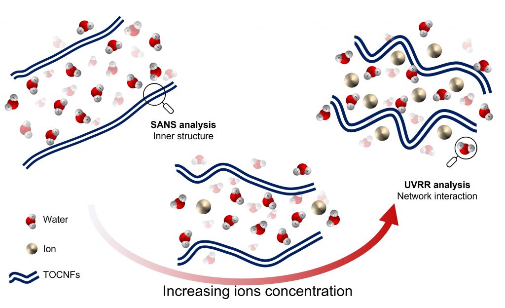
To produce nanofibrils, cellulose often undergoes to a process of oxidation catalysed by TEMPO (2,2,6,6-tetramethylpiperidine 1-oxyl). But how do these nanofibrils, and the derived hydrogels, behave in the cellular environment? To understand it, Dr Arianna Rossetti, Dr Andrea Fiorati (Politecnico di Milano) and colleagues investigated their inner structure as a function of both nanofibrils and ions (Ca2+ and Mg2+) concentration, at three different pH values. Analyses were conducted using a combination of small-angle neutron scattering (SANS) and UV Resonant Raman scattering techniques, available respectively at the Hungarian and Italian CERIC Partner Facilities.
These two complementary techniques allowed researchers to perform a comprehensive structural analysis of TEMPO-oxidized cellulose nanofibrils hydrogels at a nano and molecular scale, in different conditions: this step crucial to understand how these systems interact when they are immersed in a cellular environment, paving the way for the future development of biomedical applications.
ORIGINAL ARTICLE:


