

Synchrotron radiation-based X-ray imaging to improve knowledge about female reproductive system
|Developing an approach to investigate at high spatial definition the quality and state of ovary structures would play an important role in fertility studies and in clinical practice. In this context, a promising tool is microtomography, which allows to perform 3-D reconstruction of entire tissues and organs, through sections with up to single-cell level resolution.
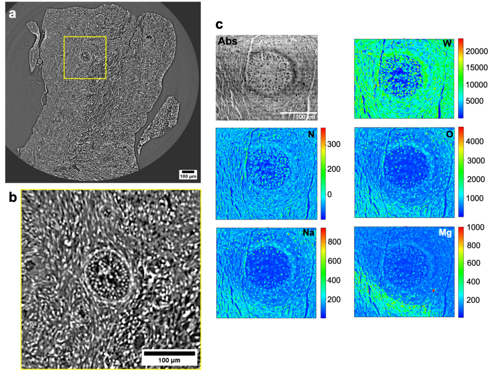
In this context, the use of contrast agents can improve the visualization of internal structures in ovary tissues, which have low radiopacity. Dr. Alessandra Gianoncelli (Elettra Sincrotrone Trieste), Dr. Lorella Pascolo (IRCCS Burlo Garofolo Hospital for Mothers and Children, Trieste) and colleagues applied four different staining protocols – based on iodine or tungsten containing agents and performed at different energies – to study in-vitro bovine ovary tissue morphology, using synchrotron radiation propagation phase-contrast microtomography (microCT). Using two synchrotron radiation available at two sites, including the SYnchrotron Radiation for MEdical Physics (SYRMEP) beamline located at the CERIC Italian partner facility in Elettra Sincrotrone Trieste, scientists discovered that tungsten-based agents allow the better identification of large follicular structures, while Iodine ones permit to define smaller intrafollicular features. The analyses were complemented by X-ray Fluorescence mapping on 2D sections at TwinMic beamline, showing that the tungsten-based agent has a higher penetration in this type of tissues.
These analyses, which have the advantage of not damaging the tested samples, could be a powerful fool to enhance our knowledge of the folliculogenesis process, having a profound impact on reproductive medicine, specifically concerning fertility preservation options for prepubertal girls with malignant tumors.
ORIGINAL ARTICLE:
