MeV Time-of-flight Secondary Ion Mass Spectrometry (MeV TOF-SIMS)
Ruđer Bošković Institute in Zagreb
For maintenance reasons the ion beam Croatian facilty at the Ruđer Bošković Institute is partially operational. Currently, the MeV Time-of-flight Secondary Ion Mass Spectrometry (MeV TOF-SIMS) is not available. Please check out the other instruments offering limited access at the Ruđer Bošković Institute at the webpage Open Access Offer
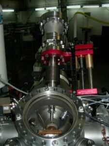
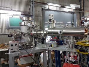
MeV Time-of-flight Secondary Ion Mass Spectrometry (MeV TOF-SIMS) is an advanced ion beam analysis technique designed for the chemical and molecular characterization of organic materials. This method relies on the interaction of energetic heavy ions with the sample, primarily through electronic energy loss. Due to the “soft” nature of this interaction, molecules are desorbed largely intact with minimal fragmentation, making MeV TOF-SIMS highly suitable for molecular identification (detecting molecules up to 1000 amu), with application in the field of biology, forensic, cultural heritage, etc. As the desorbed molecules possess energies around 10 eV, this technique is highly surface-sensitive, analyzing only the topmost layer of the sample.
At Ruđer Bošković Institute, we offer two types of MeV TOF-SIMS spectrometers for molecular imaging of organic materials:
1. Linear TOF-SIMS at Ion Microbeam Chamber

Technical specifications:
This system utilizes heavy ion beams (such as O, Si, Cl) with energies around 10 MeV to desorb molecules from the surface of organic samples. These desorbed molecules are ionized upon interaction with the sample and then accelerated towards the spectrometer using a ±5 kV voltage differential. The molecule’s mass is determined by measuring its time of flight (TOF) in a linear TOF spectrometer.
Connected to a nuclear ion microbeam, the instrument can focus ions down to the micrometer range, enabling 2D molecular imaging. It can operate in two modes:
- Pulsed mode: Uses a chopped primary ion beam for the TOF start signal, providing lateral resolution of ~10 µm.
- Continuous mode: A continuous ion beam is used with the TOF start signal generated by a particle detector positioned behind the sample. This offers higher lateral resolution (~300 nm) but requires the sample to be transparent to 10 MeV ions.
The mass resolution is M/ΔM ≈ 500 for M ≈ 100 Da, and molecules up to 1000 amu can be analyzed. The system supports sample areas up to 1×1 mm² in a single scan.
Sample Requirements:
- Samples should be placed on a conductive substrate.
- For high-resolution 2D imaging (sub-1 µm), samples should be very thin (~5 µm) and mounted on a Si3N4 window with a ~10 nm gold coating.
- Care should be taken to avoid surface contamination, and chemical fixatives or plastic tools should be avoided. Standard preparation methods such as fast-freezing in LN2, cryotome sectioning, and freeze-drying are recommended.
More details can be found here.
2. Reflectron-type TOF-SIMS Spectrometer with Collimated Ion Beam

Technical specifications:
This system operates using heavy ion beams (such as Cu, I, Au) with energies in the tens of MeV. The ion beam is focused to a micrometer-sized spot (~5 µm) using a collimator with a 5 µm aperture and a thin (~5 nm) carbon foil, achieving a lateral resolution of approximately 10 µm.
The mass analysis is performed using a TOF Reflectron analyzer, and 2D molecular imaging is achieved by moving the sample with a precise 4D piezo stage. The system supports two measurement modes:
- Transmission mode: Suitable for thin samples, with a PiN diode placed behind the target to generate the TOF start signal.
- Pulsed extraction mode: Suitable for thick samples, using secondary electrons emitted from the collimator’s carbon foil for the TOF start signal.
The mass resolution is M/ΔM ≈ 2500 for M ≈ 600 Da, and molecules up to 1000 amu can be analyzed. Sample areas up to 20×20 mm² can be analyzed in one scan.
Sample Requirements:
- Samples should be mounted on a conductive substrate.
- For transmission mode, samples should be very thin (~5 µm) and mounted on a Si3N4 window with a ~10 nm gold coating.
- Care should be taken to avoid surface contamination, and chemical fixatives or plastic tools should be avoided. Surface contamination must be avoided during preparation and handling. Use of standard fast-freezing, cryotome sectioning, and freeze-drying procedures is recommended.
More details can be found here.
As in the case of MeV SIMS, sample preparation is crucial, it is highly advisable that users contact the scientist in charge of the instrument before and during sample preparation.
Contact: dr.sc. Zdravko Siketić
Email: zsiketic@irb.hr
Tel: +385 1 4571 227
-
25.08.2025
Microfabrication Laboratory


