

Offline Synchrotron Infrared Source for Spectroscopy and Imaging – Chemistry & Life Science
Elettra Sincrotrone Trieste
SISSI Bio OFF-line is the infrared vibrational spectroscopy Laboratory at Elettra, jointly operated with the synchrotron radiation on-line facility, SISSI-Bio (Birarda et al., 2022; Lupi et al., 2007). The laboratory, recently renovated thanks to the project Pathogens Readiness Platform for CERIC-ERIC upgrade (PRP@CERIC)*, is equipped with state-of-the-art instruments for infrared spectroscopy, microscopy and imaging based on glow-bar and laser sources, capable to cover the far and mid infrared regimes. Chemical details can be achieved at different scale of spatial resolution, from the macro-scale (FTIR spectroscopy) to the micro (FTIR microscopy), sub-micron (Optical Photothermal Infrared) and the nano-scale (IR nanoscopy) on bulk materials, surfaces and interfaces.
The applications of infrared vibrational analysis cover a wide range of research fields, including, among others, Life and Medical Science, Molecular Biology, Biophysics and Biochemistry, Environmental Science and Cultural Heritage. More details on the available instruments are given in the technical specifications.
Contact: Lisa Vaccari
Tel: +39 040 375 8465 (office) | +39 040 375 8567 (beamline)
Contact: Giovanni Birarda
Tel: +39 040 375 8814 (office) | +39 040 375 8567 (beamline)
Technical specifications
Fourier Transform infrared spectroscopy (FTIR spectroscopy) may be performed at SISSI-Bio OFF-line by exploiting a large variety of interferometric systems and associated accessories. The VERTEX 70v (see Figure 1) is an in-vacuum interferometer that may operate in the Mid (MIR) and Fir (FIR) regimes, by using single point DTGS/MCT and bolometric detection schemes respectively. A special technology capable to cover the FIR and MIR in a single scan, named Bruker FM, is also available. Conventional transmission measurements may be done and accessories are available for Single reflection Attenuated Total Reflection, ATR (Platinum by Bruker, Miracle by PIKE Miracle and G-ATR by Harrick Scientific), 25-reflection ATR (25-reflections by Harrick scientific), Infrared Reflection Absorption Spectroscopy accessory (A513/Q Variable angle reflection accessory by Bruker). Diamond compression cells (2 mm culet) and a diamond anvil cell (800 µm culet) are available.
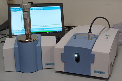
The laboratory is also equipped with the ALPHA II (See Figure 2) compact interferometer by Bruker (PRP@CERIC funded instrument). Conceived for being portable and easy to use, the ALPHA II system is equipped for ATR and reflection non-contact measurements, particularly useful for surface analysis of bulky samples.
An additional VERTEX 70 interferometer is also available.

SISSI-Bio OFF-line features two FTIR microscopes, for correlating the morphological features of a sample with its local chemistry in the MIR. The Hyperion 3000 Vis/IR microscope (see Figure 1), coupled with the vertex 70v microscope, is equipped with a single point MCT detector for single point measurements and hyperspectral mapping. It also mounts a bidimensional Focal Plane Array (FPA) detector for hyperspectral imaging (64X64 pixels). Transmission, reflection, Grazing incidence and micro-ATR sampling modalities are possible (15X, 36X objectives; micro-ATR objective; grazing incidence (GIR) objective). In addition, a FTIR-600 Linkam cell devoted to dedicated T-dependent studies (-195°C-600°C) may be exploited.
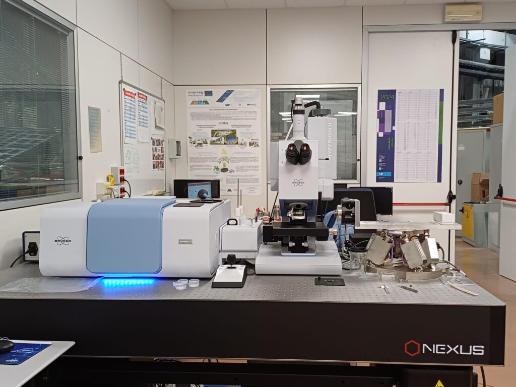
A new Bruker Hyperion II microscope coupled with the INVENIO-II interferometer (see Figure 3) has been recently installed (PRP@CERIC funded instrument). With respect to the Hyperion 3000 microscope, the Hyperion II mounts a larger FPA detector (128×128 pixels) for faster imaging or larger areas. The system is also devoted to IR tomography measurements.
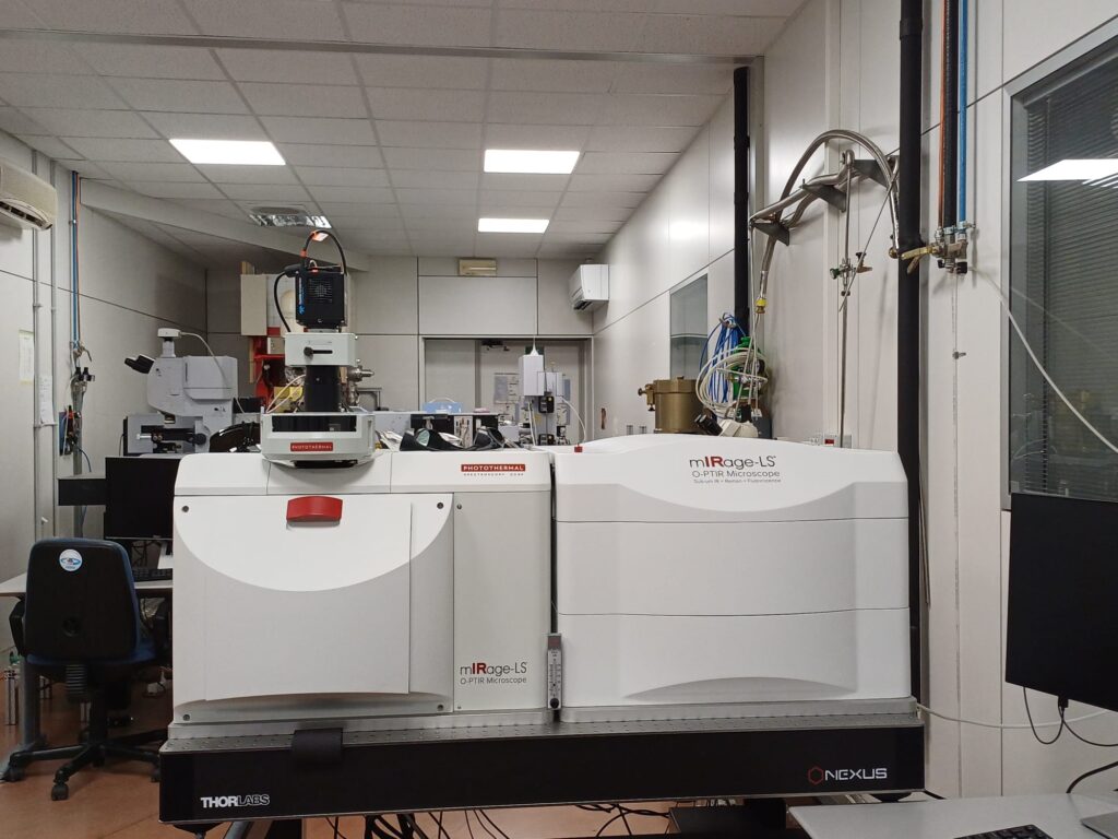
A microscopy endstation for vibrational hyperspectral mapping and imaging with sub-micrometer lateral resolution is available (see Figure 4). The former experimental station is based on the so-called optical photothermal infrared (O-PTIR) microscopy, also known as Midinfrared photothermal (MIP) microscopy (PRP@CERIC funded instrument). The O-PTIR approach may be summarized as an IR-pump/visible probe approach. It works by illuminating a sample with pulsed IR radiation, from a tunable IR quantum cascade laser (QCL). When IR light excites molecular bonds within a sample, a portion of the energy absorbed and dissipated in the sample results in localized heating that, in turn, causes a reduction of the index of refraction of the IR-heated regions. An optical probe beam, i.e. a green-laser, detects the IR-induced changes that alter the intensity and angular distribution of probe light reflected, scattered, and/or transmitted by the sample. O-PTIR microscopy is diffraction limited. However, despite conventional IR microscopes, the aforementioned limit is imposed but the probing visible laser. Considering that visible light wavelengths are on average 1 order of magnitude shorted than Mid-IR wavelengths, the achievable lateral resolution is about a factor 10 better, allowing to switch from the micron to the sub-micron scale.
In addition, O-PTIR supports simultaneous Raman microscopy: sample scattered light may be separated by a dichroic mirror: elastic scattered light is exploited for probing IR sample absorption, whereas inelastic scattered light is sent to a Raman spectrometer for the generation of Raman spectra. As such, the IR and Raman measurements are obtained at the same time, same location, and same spatial resolution. In addition, fluorescence microscopy is also combined within the same instrument.
For more details on the technique and application, the reader may refer to the following recent paper: Craig B. Prater, Mustafa Kansiz, Ji-Xin Cheng; A tutorial on optical photothermal infrared (O-PTIR) microscopy. APL Photonics 1 September 2024; 9 (9): 091101. https://doi.org/10.1063/5.0219983
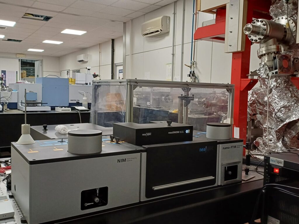
At SISSI-Bio OFF-line, CERIC-ERIC users may also perform vibrational analysis at the nanoscale exploiting the possibility offered by the near-field NeaSNOM microscope, by Attocube GmbH (see Figure 5). The equipment is suitable for surface analysis by IR scattering-type scanning near field optical microscopy (IR s-SNOM) in the 670-1800 cm-1 spectral with a DFG laser source (nano-FTIR module) and Photo Thermal Expansion imaging feature AFM-IR modality of the instrument (NIM module) with a QCL laser source.
*under the PNRR (National Recovery and Resilience Plan) under Mission 4 “Education and Research”, Component 2 “From Research to Enterprise”, Investment Line 3.1 “Fund for the creation of an integrated system of research and innovation infrastructures”,” funded by the European Union – Next Generation EU”.

-
20.02.2025
X-ray computed tomography (CT) TomoLab
-
23.01.2025
THz beamline TeraFERMI