

X-ray computed tomography (CT) TomoLab
Elettra Sincrotrone Trieste
X-ray computed tomography (CT) is a non-destructive testing method which offers an attractive opportunity for the three-dimensional (3D) insight of the inner structure of objects and materials. Originally introduced for medical diagnostics by Hounsfield in 1972, this technique has been significantly improved and adapted to be used for research and industrial applications since early 1990’s.
Due to the great technological advances in the realization of detectors and X-ray generators and to the computational power of modern computers, X-ray computed microtomography (microCT) systems, capable to reach a micrometric spatial resolution on centimetre-sized samples.
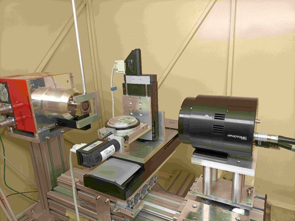
The object under investigation, without any special preparation, is placed on a rotary table between the X-ray source and the detector: from hundred to thousand radiographs (projections) are then acquired during the sample rotation (usually over 360 degrees). A dedicated software processes these projections in order to reconstruct a sequence of axial slices of the sample which, in turn, can be stacked to create a 3D dataset.
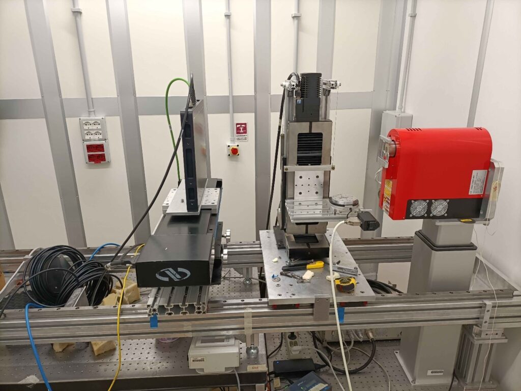
In the laboratory there are two custom systems based on conventional microfocus X-ray sources. The first one is hosted a shielded cabinet and it is dedicated to samples around 20 mm diameter and 30 mm height with a final voxel side in the range 5-15 micron. The second one is placed inside a walk-in hutch and it can acquire objects up to 200 mm diameter and 300 mm height (it will be upgraded soon) with a final voxel side in the range 10-60 micron.
In case of any specific requirement or need (e.g. bringing own set-up to condition the sample), proposers are invited to contact the laboratory team before submitting a proposal.
Contact:
Diego Dreossi
Tel: +39 040 375 8036 (office) | +39 040 375 8765 (laboratory)
Technical specifications
TOMO 1 cabinet micro-CT scanner
- Sealed microfocus X-ray tube, 40-130 kV, 39 W, focal spot size down to 5 micron
- Air cooled sCMOS, 4k x 4k pixel matrix, 13.2 μm pixel size (54 mm X 54 mm field of view)
TOMO 2 walk-in hutch micro-CT scanner
- Sealed microfocus X-ray tube, up to 150 kV, 75 W, focal spot down to 5 micron
- Air-cooled sCMOS, 2k x 2k pixel matrix, 31.2 μm pixel size (63.9 mm X 63.9 mm field of view)
- Flat panel sensor, 2192 x 1776 pixel matrix, 120 micron pixel size (263 mm x 213 mm)
Sample environment
Both systems can accommodate special devices or external equipment to create controlled experimental/physical conditions on the sample during scanning (e.g. for temperature-controlled testing, stress-strain monitoring, fluid injection). It is also possible to manage additional detectors in a coordinated manner with the rest of the instrumentation.
Detailed information can be found on the beamline’s main homepage.
-
23.01.2025
THz beamline TeraFERMI
-
15.01.2025
ATHOS Triple-Axis Spectrometer