PhD Scholarships
- PhD Scholarships
- Highlights
- Scientific publications
- In-house research
- International collaborations
- Resources
- Guidelines
- Procurement
PhD Scholarships
THE OBJECTIVE OF CERIC-ERIC PhD PROGRAMME ACTIVITY IS TO FURTHER THE INTEGRATION OF THE PARTNER FACILITIES AND TO CONTRIBUTE TO EXCELLENT SCIENCE.
CERIC PhDs Guidelines
These Guidelines aim to ensure that the research carried out by the PhD students properly represents the role and identity of CERIC-ERIC and acknowledges the support received. In addition, they aim to increase the visibility of the PhD research by communicating about its progress and disseminating its results.
CONTACT: phd@ceric-eric.eu
Naming CERIC-ERIC
When naming CERIC for the first time, in any written communication or in a speech, use the following: name, acronym: Central European Research Infrastructure Consortium, CERIC-ERIC. It is then possible to use only CERIC later in the text/speech. In all oral presentations, the acronym is pronounced [serik-erik].
Naming the partner facilities in CERIC
Whenever you need to mention a partner in your project that is a CERIC partner facility, or whenever you refer to any other CERIC facility, please use the naming in the forms as listed below:
Austria: Austrian CERIC partner facility at the Technical University Graz and in Trieste.
Croatia: Croatian CERIC facility at the Ruđer Bošković Institute in Zagreb.
Czech Republic: Czech CERIC partner facility at the Charles University in Prague and in Trieste.
Hungary: Hungarian CERIC partner facility of the Centre for Energy Research (EK) in Budapest.
Italy: Italian CERIC partner facility at Elettra Sincrotrone Trieste.
Poland: Polish CERIC partner facility at the National Synchrotron Radiation Centre SOLARIS in Krakow.
Romania: Romanian CERIC partner facility at the National Institute of Material Physics (NIMP) in Magurele.
Slovenia: Slovenian CERIC partner facility at the National Institute of Chemistry in Ljubljana
CERIC acknowledgement
When introducing CERIC in written statements/communications, the following text (CERIC mission) should be always included:
“CERIC is a research infrastructure integrating and providing open access to facilities in Europe to help science and industry advance in the fields of materials, biomaterials and nanotechnology”.
When introducing CERIC in written statements/communications, the following text (CERIC mission) should be always included:
“This work is part of the PhD “insert your topic” supported by CERIC-ERIC”.
Presentation to ISTAC
CERIC will ask to present your research to CERIC’s ISTAC (International Scientific and Technical Advisory Committee), either during dedicated meetings or in scientific events organised by CERIC.
Please remember to let us know about any relevant event that could be of interest to us (e.g. prize) and to send us a copy of any reports you produce during your PhD.
Confidential Information
Consider private and confidential information received from CERIC-ERIC related to obtaining the PhD qualification.
Always include the CERIC logo in your posters, presentations, etc. related to your project.
Either when you use the CERIC logo alone, or when you associate it with logos of other institutions, please refer to and apply the Guidelines for a correct use of the CERIC logo.
- For all presentations related to CERIC internal research projects, please use this .ppt template
- For the production of project-related documents/reports, please use this headed paper template
In your presentations, whenever possible, include a slide introducing CERIC. Download it here.
CERIC Press & Communication office can provide support for the following activities, if they are aimed at increasing the visibility of your research, as well as of CERIC and its facilities:
- Draft, publication and promotion of scientific highlights and articles on your research results;
- Draft/distribution of press releases to Italian and European media (radio, tv, newspapers);
- Highlight of your research results in CERIC quarterly newsletter;
- Realization of audio and video interviews;
- Promotion of your research on CERIC social media channels (Follow us on Twitter, Linkedin and Youtube, and tag CERIC account when posting about your research);
- Supply of templates, pictures and other communication material, upon request.
We would like to keep you informed about the activities, opportunities and results of CERIC through our quarterly newsletter. Moreover, we will inform you about initiatives, courses, webinars, etc. that could be useful for you and your research activity.
WE HELP YOU IN PROMOTING YOUR PhD PROJECT’S EVENTS AND INITIATIVES!
Contact us if you would like to receive support in promoting your project’s initiatives, such as courses, events, workshops, results, etc.
DISSEMINATE YOUR PROJECT’S RESULTS TO THE WIDER PUBLIC!
Media relations shall be handled in coordination with the CERIC press office:
Contact us once your manuscript is accepted (the earlier, the better!), for the CERIC press office to evaluate eventual outreach activities and draft a press release to be distributed to the media as soon as the scientific paper is published.
Contact us if you’d like to promote single initiatives related to your research (courses, events, workshops, results, etc.).
PhDs Theses
CERIC supported the research of several PhD students (thanks to both direct support and the participation of in-house projects), whose experiments and results were published as part of their dissertations.
- Zingaro F., Advanced approaches to investigate immune and reproductive toxicity of nanoplastics, 2024
- Morasso S., Integrative approach in drug discovery against SARS-CoV2: a biochemical and biophysical study on the Papain-like protease, 2024, link
- Moradmand S., Towards fabrication of 2D out-of-plane functional supramolecular nanoarchitectures, 2023
- Wechsler D., Porphyrin adsorption and reactivity on oxide surfaces, 2021, link
- Kosto Y., Study of cerium oxide thin films for biosensing applications, 2021, link
- Bercha S., Model systems with the use of CeO2 for bioapplication, 2020, link
- Bogar M., Operando SAXS investigation for Polymer Electrolyte Fuel Cells, 2020
- Wituschek A., Phase-modulated coherent time-domain spectroscopy in the extreme ultraviolet regime, 2020, link
- Zalduendo M.M., Composite materials based on mesoporous thin films and metallic nanoparticles: synthesis, characterisation and design of optical sensors, 2020
- Bottari C., Solvation effects of ionic liquid/water mixtures on biomolecules, 2020, link
- De Angelis D., Graphene-based interfaces as tuneable support for metal oxide nanoparticles, 2019, link
- Lionello D., Study of mechanical and tribological properties of nanostructured materials, 2018
- Kettner M., Reactivity of transition metals – influence of the degree of oxidation of active substrate, 2017, link
- Beranova K., Study of dependence of the metal-oxide electron structure on the reactivity of these systems, 2015, link
CERIC PhDs (ongoing)
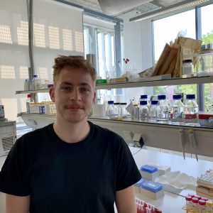
Bio:
Simon Aleksič was born on April 19, 1995, in Slovenia. He received his B.Sc. and M.Sc. degrees in Biochemistry in 2017 and 2020, respectively, at the Faculty of Chemistry and Chemical Technology, University of Ljubljana. Since 2020, he is employed at the Slovenian NMR Centre as a Ph.D. student, supported by the CERIC-ERIC consortium, with a research focus on the effect of oxidative stress on guanine-rich genomic regions using high-resolution NMR spectroscopy.
Title of research:
Effect of oxidative stress on guanine rich genomic regions
Abstract:
Guanine-rich nucleic acids can adopt noncanonical structures, called G-quadruplexes. Potential G-quadruplex forming sequences are predominantly found in the promoters and telomeric regions of genomes and studies show that their formation can affect important cellular mechanisms, such as gene expression and telomere length regulation. During periods of oxidative stress in the cell, reactive oxygen species can chemically damage biomacromolecules. Among the four common nucleobases, guanine is the most susceptible to oxidation. G-quadruplex forming sequences are therefore potential sites of intensified guanine oxidation. Oxidated lesions feature altered hydrogen bonding abilities, which can impact the folding, structure and stability of nucleic acids. Using high-resolution NMR methods and complementary techniques, we will determine how oxidation of guanine impacts the structure of guanine-rich nucleic acids, originating from functionally important genomic regions.

Bio:
Alessandro Alleva was born in Legnano in 1996. He studied Materials Engineering and Nanotechnology at Politecnico di Milano, completing there both his Bachelor’s and Master’s studies. During his academic career, he spent six months at L’École normale supérieure de Lyon as part of the Erasmus+ project. He carried on his Master’s thesis work in IMEC (Belgium), focusing on thin films deposition and characterization for high efficiency photovoltaics applications. Currently, he is enrolled as a PhD student in Politecnico di Milano and his work is centered on Li an post-Li battery technologies.
Title of research:
Morphochemical and structural changes of electrodes and electrolytes in all-ceramic solid-state lithium batteries
Abstract:
Renewable energy sources could replace hydrocarbons, but sustainability imposes integration with reliable and efficient electrochemical energy storage facilities, among which Li-based systems currently play a pivotal role. Commercial Li-ion batteries generally employ liquid electrolytes, which show poor stability in contact with the electrodes. In this scenario, solid-state batteries, with both Li and post-Li technologies, are a promising solution for next-generation energy storage, owing to their high energy density, light weight and high safety compared to the current technology based on flammable liquid organic electrolytes. This work will be centered around electrochemical investigations, soft X-ray absorption microspectroscopy and X-ray fluorescence for the assessment of the morphochemical evolution of the battery materials during operation, especially tracking the chemical state and space distribution of the electrodic metals at the electrode-electrolyte interface.

Bio:
Anastasiia Deineko, PhD student at the Department of Surface and Plasma Science, Faculty of Mathematics and Physics, Charles University, Prague, Czech Republic. My research explores cerium oxide as an electrode material for the detection of biomolecules, in particular glucose and urea. It combines surface analysis, which is performed at the Material Science Beamline of Elettra Sinchrotrone, Trieste, and electrochemical measurements, which are carried out at the department in Prague.
Title:
Development of ceria based electrochemical sensors for biomolecule detection
Abstract:
Cerium oxide is a promising electrode material as it possesses enzymatic properties for the detection and determination of various biologically important molecules. We were characterizing polycrystalline cerium oxide prepared on a glassy carbon substrate as an electrode material for glucose and urea detection. Target biomolecules are analyzed by photoemission spectroscopies (XPS, SRPES, RPES, NEXAFS) to determine whether the molecules bond to the surface in the absence of electrochemical potential and if so, the nature of the chemical bond – physisorption or chemisorption. In parallel, electrochemical measurements are carried out to determine detection limits and linearity of response. Sample characterization is performed under vacuum (first step) then it will be done under increased water pressure to determine the effect of water on the adsorption of the target molecules. Based on the results obtained, exploratory experiments will be carried out to determine whether the performance of the sensors (detection limit, linearity, stability) can be improved by doping the nanostructures, for example.

Bio:
Matej Gabrijelčič is a Researcher PhD Student in Physics who works in the field of solid state NMR and modern battery systems at the National Institute of Chemistry in Ljubljana (Slovenia). Matej holds a Bachelor’s Degree in Physics and a Master’s Degree in Physics Education from the University of Ljubljana. Matej has 4 years of experience in the automotive industry, especially R&D and laboratory for testing and validation. He is motivated in constant improvement in natural sciences, combined with learning about soft skills. He is a certified NLP Master Coach from International NLP Trainers Association. In his free time, Matej likes to practice ju-jitsu and improvisation theatre.
Project title:
Unravelling the electrochemical mechanisms of battery degradation by operando NMR and X ray absorption spectroscopy
Abstract:
The goal of the proposed PhD study is the implementation of operando NMR spectroscopy of batteries and related materials at SloNMR@NIC. The PhD student will install, modify and test the necessary equipment and then use it to study the degradation of batteries. The operando NMR measurements will be combined by the operando XAS (performed at Elettra) and XRD measurements (performed at NIC or Elettra). The aim of the measurements will be to clarify the degradation mechanisms in the bulk (XAS, XRD and NMR) and at the interfaces (NMR). Such a combined study will provide the necessary data for the evaluation of battery quality and expected lifetime.

Bio:
Athira lekshmi M S is a PhD student at Charles University, Prague working under the supervision of Dr. Ivan Khalakhan in the Nanomaterials group of the Department of Surface and Plasma physics. She completed her Masters in Physics in 2020 from the Central University of Tamil Nadu, India with outstanding grades. At the Central University of Tamil Nadu, she worked on Experimental Condensedmatter Physics under the supervision of Dr. K C Sekhar for her Master’s Research project whichfocused on studying Energy storage capacitor applications of Relaxor ferroelectrics. Currently, she is working on monitoring the relationship between morphological, structural and compositional degradation of the catalyst and operating conditions of fuel cells.
Project title:
Unraveling deterioration of fuel cell catalysts
Abstract:
The catalytic layers (Pt and Pt-based bimetallic alloys) will be prepared using the magnetron sputtering technique for application in the cathode of proton exchange membrane fuel cells (PEMFCs). Deposited catalysts will be characterized by electron microscopy (SEM, TEM), photoelectron spectroscopy (XPS) and X-ray diffraction (XRD). The identical catalysts will be then characterized with the same techniques after stability tests performed in an electrochemical cell where the PEMFCs operating conditions will be recreated. The in situ characterization of catalyst directly during simulated operational conditions will be studied by employing in-situ electrochemical atomic force microscopy (EC-AFM) and rotating disc electrode (RDE). The goal will be to establish the connections between morphological, structural and compositional degradation of a catalyst and operation conditions of the fuel cell.
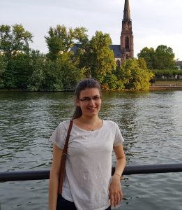
Bio:
Catalina-Gabriela Mihalcea was born in Bucharest, Romania and is a Research Assistant at the National Institute of Materials Physics, Magurele, Romania. She is a PhD student in Condensed Matter Physics at the University of Bucharest, Faculty of Physics. Her research field is focused on Analytical Transmission Electron Microscopy techniques and X-ray Diffraction. She received the Bachelor’s Degree in Physics from the University of Bucharest, Faculty of Physics, in 2018 and the Master’s Degree in “Physics of Advanced Materials and Nanostructures” from the University of Bucharest, Faculty of Physics, in 2020.
Project title:
Nanostructured materials for gas sensing: correlation between functional, electronic and 3D microstructural properties
Abstract:
This PhD research is focused on “Nanostructured materials for gas sensing: correlations between functional, electronic and microstructural properties”.
Gas sensors have been developed for applications in different areas, such as domestic safety, environmental monitoring, automotive, spacecraft or medical diagnoses. Solid-state gas sensors based on semiconducting metal oxides (SnO2, ZnO, TiO2, In2O3, etc.) are currently the most frequently used. The main issues that are still currently under debate are related to their selective sensitivity, detection limit, reliability, influence of the water vapor on the gas sensing performance, etc. New materials and/or synthesis routes are currently approached in order to improve their characteristics. (Sens. and Act. B 204, 250–272, 2014). The correct understanding of the physical-chemical mechanisms underlying the detection process may be accomplished by applying appropriate microstructural (XRD, SEM, HRTEM, STEM) and spectroscopic (XPS, EDS, EELS) investigation techniques along with the specific measurements of the electrical properties under controlled atmosphere to evaluate the global and local morphological, structural and chemical properties of the approached material. However, a serious obstacle in getting a reliable correlation between the measured electrical properties and especially the local chemical information results from the fact that the corresponding measurements are usually performed in different experimental conditions (Sensors 18, 3544, 2018). Namely, XPS and TEM studies are performed under vacuum or ultra-vacuum conditions which means far from the real operational conditions. For this reason, in-situ and operando techniques are needed in order to analyze the fine mechanisms at the electronic and chemical level.
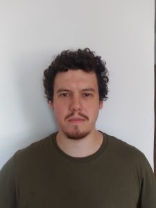
Bio:
He is a secondyear doctoral student at the University of Bucharest in Romania, at the Faculty of Physics, in the Condensed Matter Physics program. His research activity concerns the physical properties – such as structure, optical, electrical properties – in advanced materials, involving various transmission electron microscopy techniques (TEM/STEM) and analytical methods such as EDS (energy dispersive X-ray spectroscopy) and EELS (electron energy loss spectroscopy).
Project title:
From crystal structure to functionality: tailoring the strain driven physical properties of materials
Abstract:
The topic of the thesis concerns the influence of structural information (strain, defects) on electrical properties (electrical polarization, potential barrier, work function) of ferroelectic thin films by correlating combining advanced microstructural and spectroscopic techniques with electrical measurements. Ferroelectric oxides, such as Pb(Zr,Ti)O3, are useful for electronic and photonic devices because of their ability to retain two stable polarization states. Transmission electron microscopy proved to be a powerful technique for studying the crystalline structure and chemical composition at the interface (e.g. electrodes), at domain walls and allows the visualization of the polarization field in ferroelectric heterostructures. Other high-resolution spectroscopic techniques like, especially X-ray photoelectron spectroscopy (XPS), but also electron energy loss spectroscopy (EELS) can provide important information on the influence of strain on the electronic properties.
Bio:
Sebastian Rücker was born in Steyr, Austria, in 1994. From 2014 on he studied chemistry and biology as teachers training at the Karl-Franzens Universität Graz where he graduated in 2021. For his Diploma he worked on nickel-based paddlewheel complexes. Due to this experience he developed a great interest in research and decided to apply for the PhD program at the Technische Universität Graz in 2022. There he is now pursuing his PhD degree with great enthusiasm as part of a very international team.
Project title:
Continuous flow synthesis of MOF biocomposites
Abstract:
Metal-organic frameworks (MOFs) are a class of materials that have shown capabilities as carriers for the delivery of biopharmaceuticals or for the protection of proteins from hostile environments. Despite the current progress in the integration of different proteins in/on MOFs, high enzyme activities, tuneable particle sizes, reaction time control and scalability could only be partially achieved with the bulk synthesis approach. This PhD project aims to establish new fabrication methods for the integration of biomolecules in different MOF materials. A customized in flow system will be used to control the structural and functional properties of the MOF composites. Small angle X-ray scattering (SAXS) analysis will help to correlate the synthesis variables with the final performance of bioactive MOF composites.
Bio:
Project title: Hydration of bio-molecules by combined use of synchrotron-based UV Resonance Raman and Neutron Scattering experiments
Abstract:
CERIC PhDs (concluded)

Bio:
She studied Biomedical Engineering for her B.Sc. and undertook an M.Sc. in Bionanotechnology, both at the Polytechnic of Turin, and now she is a 1st year Ph.D. student in the Nanotechnology program at the University of Trieste. In her master thesis project, which was part of an exchange period as a visiting student at the Uppsala University, she worked on the characterization at the single-particle level of small extracellular vesicles, by using Atomic-Force Microscopy and Fluorescence Microscopy, for cancer diagnosis. This experience showed her the different ways in which fundamental and applied research may interact and enriched her passion on how the physical and biological worlds can works in synergy for the development of important biomedical applications. To follow that line, she is currently working on a multidisciplinary Ph.D. project that aims to understand the role played by small extracellular vesicles in cell-cell communication mechanisms, with an eye to cancer applications
Project title:
Mechanisms of Extracellular Vesicles (EVs)internalization by cells
Abstract:
Small extracellular vesicles (sEVs) are nanometer-sized vesicles (30-200 nm) that are released from almost all types of cells both in pathological and physiological conditions. These biological nanoparticles have shown potential for cancer diagnostics/therapeutics as by travelling in body fluids (e.g., blood, saliva) they carry “biological information” between near and far cells. However, the complexity of EVs origin, their composition, and nanometric size, make them a challenge to be analysed by many available bioanalytical techniques. Indeed, EVs are heterogenous nano-carriers with complex cargoes (proteins, lipids, and nucleic acids, etc.), whose population mostly changes in accordance with the type of parental cells and the specific physiological or pathological conditions at the moment of their packaging and secretion. Cell type specificity of EVs biogenesis, release, and uptake is still debated, as well as the involved targeting cell receptors and internalization pathways (e.g., cell membrane fusion and endocytosis).
Herein, we propose a nano/microscopic-based approach to investigate structure-function correlations of EVs from two different breast cancer cell lines with different aggressiveness, with lipid model membrane systems of increasing complexity. As a starting point, AFM imaging was and will be performed by using lipid bilayers of variably composition, in different environmental conditions. Preliminary vesicle-membrane interaction analyses were carried out with sEVs isolated from human umbilical cord-derived mesenchymal stromal cells (hUC-MSCs) as a reference sample. Results obtained revealed various interaction mechanisms of EVs with lipid model membranes depending on the salt concentration and preferentially with liquid-ordered raft-like lipid domains. These findings allow a better understanding of the EV uptake mechanisms by recipient cells and the most relevant parameters involved in the EV selective targeting in tumor microenvironments.

Bio:
Antonio Longo (15/07/1995 – Battipaglia (SA)) defines himself as a person in love with the mechanisms at the basis of music and life. Giving priority to only one of the two, he studied biological sciences at the University of Salerno and functional genomics at the University of Trieste, where he achieved the master degree cum laude in 2019, presenting a master thesis entitled “Structural characterization of the C-terminal region of the RTEL1 helicase”. In 2020 he obtained a joint fellowship ICGEB – Elettra, joining the Structural Biology laboratory at Elettra (led by prof. Silvia Onesti) and the Molecular Pathology laboratory at ICGEB (led by prof. Emanuele Buratti), investigating the biochemical properties of the human RECQ4 helicase.The same year (2020) he started his INTEGRA-CERIC PhD program in Chemistry (University of Trieste), in the Structural Biology Laboratory at Elettra Sincrotrone Trieste.
Project title:
Structural and functional analysis of helicases involved in genome maintenance
Abstract:
Helicases are molecular motors able to unwind nucleic acids structures by using energy from ATP hydrolysis. Among the Superfamily 2 helicases, the RecQ family and the FeS family, conserved from bacteria to higher eukaryotes, are characterized by a wider range of nucleic acid substrates, including D-loops, R-loops, G-quadruplexes, triple helices, Holliday junctions, etc. RecQ helicases are essential in preserving the genomic stability of cells, by performing a broad variety of functions at the interface of DNA replication, recombination and repair. In humans, there are five RecQ helicases, of which three of them cause rare genetic syndromes when mutated. The FeS helicases are characterized by the presence of a domain that binds an iron-sulfur (FeS) cluster which seems to be involved in nucleic acid unwinding. Defects in every of the FeS helicases cause genetic syndromes and cancer predisposition. Some structural information is available for human RecQ helicases and none for Fe-S helicases. Their involvement in many severe diseases highlights the relevance of additional biochemical and structural information, to better understand their function in the cell, the role of the “non-helicase” domains (often essential for viability) and to elucidate their interactome, which are tightly regulated in concert with the cell-cycle. In this context, the aim of this PhD project is to characterize structurally and functionally key helicases for genome maintenance. For each target helicase the project will focus around 3 major activities: 1) the production of the recombinant protein and its mutants; 2) the biochemical characterization of these proteins; 3) the use of all these proteins for structural investigations using X-ray crystallography, small-angle X-ray scattering and cryo-electron microscopy.

Bio:
CarloAlberto is a PhD student in chemistry working at Elettra Sincrotrone Trieste. His background covers both chemistry and materials science. During his Bachelor’s degree, he synthesized and studied up-converting luminescent nanoparticles. Moving to the Master’s program he decided to grow and study magnetic ultrathin-films at the Nanospectroscopy beamline, where he is pursuing his PhD degree at the moment. He is part of an Italian- Swedish group working on ultrafast dynamics in condensed matter focusing on magnetism. Besides being in the lab he likes to do handy activities like cultivation, gardening, beekeeping.
Title of research:
Interlayer magnetic coupling mediated by Dirac materials
Abstract:
Targets of this research are understanding and control of the macroscopic magnetic response of ferromagnetic materials when they interact with two-dimensional materials such as graphene or others Dirac materials. The main goal is to build up a synthetic antiferromagnet composed of a stack of ferromagnetic layers separated by a two- dimensional non-magnetic spacer, hence observing how the non-magnetic spacer modifies the magnetic response of the system. The research activity is performed at the Nanospectroscopy beamline at Elettra Sincrotrone Trieste, giving the possibility to prepare samples in situ and to obtain structural, chemical and magnetic information in a laterally-resolved manner.
Project title: Recovery and characterization of layered oxides materials from spent batteries: a step forward towards sustainability
Abstract:
Project title:
Integrated structural analysis of human USPs: a novel family of drug gable targets

Bio:
Lorenzo D’Amico was born in Trieste in 1994. After graduation in 2014 in Material Engineering at the University of Trieste, he decided to enrol in the master program of Biomedical Engineering. During his master he had the opportunity to join the Erasmus program and spend 6 months abroad at the Eindhoven University of Technology, where he wrote his master thesis. Thanks to this experience he developed a huge interest in research and decided to apply for the PhD program after my graduation in 2019. In August 2020 he started an internship at SYRMEP beamline (Elettra Synchrotron), where he is currently working, during which I had my first meeting with the world of research. In November 2020 I started my PhD. So far, I am very fulfilled with my choice and I look forward to attempting new challenges.
Project title:
Imaging and characterization of fibrotic tissues
Abstract:
Fibrosis is a general term for diseases that lead to an increased deposition of fibers, which in turn changes mechanical properties such as stiffness of an organ. This may affect the function of the organ like in the case of lung fibrosis the lung function. So far fibrosis has been studied very organ specific, but the underlying processes are most likely more general. It is important to underline that If the remodeling of the organ is still ongoing typically by an inflammatory process it can be suppressed with treatment. Therefore, early diagnosis of a fibrotic process and a treatment that reduces or cancels its development is of great interest. Unfortunately, little is known about the type of fibers deposited, their orientation and the underlying pathomechanism.
Therefore, the aim of this PhD thesis is to develop a multi-parametric analysis exploiting several techniques such as phase contrast microCT, histology, NIR-spectroscopy, AFM and SAXS that allows a comprehensive characterization of fibrosis.
To establish this pipeline tissue of commonly used mouse fibrosis models such as the Bleomycin lung fibrosis model will be used. Later on we plan to study patient tissue biopsies with the same pipeline. The resulting data should then be clustered using unsupervised machine learning strategies to identify potential subgroups of fibrosis.
This thesis is committed to answer the following questions:
Does the proposed combination of analytic techniques allow for the identification of fibrosis in general?
Do subtypes of fibrosis exist?
Which are the main parameters driving the decision? (Can the complexity of the analysis be reduced?)
Which subtype of fibrosis resembles the fibrosis in the typically applied animal models?
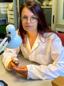
Bio:
Clarissa Dominici is a naturalist with an education on prehistoric ecology, palaeontology and human evolution. As a zooarchaeologist, she studied faunal assemblages coming from the Upper Palaeolithic sequences of Grotta Paglicci (Apulia, Italy) and Grotta della Cala (Campania). She is part of the RU of Prehistory and Anthropology of the University of Siena with a role of responsibility for the Middle Palaeolithic excavation fieldwork at Grotta dei Santi (Tuscany) and Riparo L’Oscurusciuto (Apulia).
Project title:
Hunting technologies during the Middle and Upper Palaeolithic in Central-Southern Italy – Chemical and morphological analyses on lithic and bone implements
Abstract:
Her PhD project is aimed at the reconstruction of hunting strategies of Neandertals and Modern Humans in southern Italy, through the chemical characterisation of archaeological residues coming from lithic and bone implements thought to have been used as projectiles, in addition to impact fractures analysis. The integration of multiple cutting-edge techniques will allow the discrimination between different substances on artefacts, e.g. organic traces of animal and/or vegetal origin, mixtures of different compounds used for hafting or toxic essences used as poison. To allow a comparison between human adaptation in different environmental conditions, materials from four archaeological sites are being studied: Grotta della Cala and Grotta di Castelcivita in Campania and Grotta Paglicci and Riparo L’Oscurusciuto in Apulia. The project is a joint initiative of the Department of Physical Science, Earth and Environment of the University of Siena and the Chemical and Life Sciences branch of the SISSI beamline at Elettra Sincrotrone, and is performed in collaboration with TwinMic@Elettra, SYRMEP@Elettra and LIBI@RBI.
Bio:
Martina Zangari was born in Varese in 1994. In 2017 she obtained the Bachelor’s degree in Life Science and in 2019 she graduated in Molecular and Industrial Biotechnology. Currently, she attends the 1st year PhD course at University of Trieste with CERIC-ERIC scholarship. She works in Electrophysiology and Biophysics Lab in Department of Life science at UniTS and in Sissi-Beamline at Elettra Synchrotron.
Project title:
Analysis of the changes induced by asbestos fibers on the structure of absorbed proteins and lung tissue architecture
Abstract:
In the past century asbestos, a natural mineral, was used because of its resistance and low cost (1). Respiratory exposure to asbestos fibers is a hazard to human health, because their inhalation causes cell proliferation and cells oxidative stress. A key process in pathology development is the interaction of fiber with proteins: it can trigger the entry in the cytosol, can induce cytoskeleton modification and apoptosis. Many proteins are able to bind to the asbestos fibers forming asbestos body (AB) (2). The goal of this project is to unravel the mechanism of protein binding to asbestos fibers and their consequent structural and functional modifications. First, biochemical profile modifications will be evaluated with a portfolio of techniques that exploits synchrotron radiation IR and X-ray sources (micro and nano FTIR, XRF and X-Ray Microscopies), as well as electron and protons beams (HR- TEM). Then, electrophysiology will be used exploiting the two-electrode voltage clamp on Xenopus oocytes and human cells treated with asbestos fiber to highlight the role of fibers composition on protein misfolding and toxicity.
References:
(1) Nagai H., Ishihara T., Lee WH., Ohara H., Okazaki Y., Okawa K., Toyokuni S. (2011). Asbestos surface provides a niche foroxidative modification. Cancer Science.102(12): 2118-25. DOI
(2) Bardelli F., Giulia Veronesi G., Capella S., Bellis D., Charlet L., Cedola A. & Elena Belluso E. (2016). New insights on the biomineralisation process developing in human lungs around inhaled asbestos fibres. Scientific Reports. 7:44862 DOI
Bio:
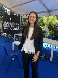
Federica Zingaro graduated in Pharmacy (2015-2020) at University of Bari Aldo Moro, Italy, with a thesis on “Self-assembled amphiphilic cyclodextrins as anticancer drug carriers for the treatment of brain tumors”, supported by the Global Thesis project between the Pharmaceutical Technology research group of Bari and the Laboratoire de Glycochimie in Amiens, France. In November 2020, she joined the PhD program in Nanotechnology at University of Trieste, to acquire advanced analytical skills to be applied in the biological fields.
Project title:
Effects of particulate and endocrine-disrupting metals on fertility
Abstract:
Long-term exposition of the reproductive system to environmental pollutants is raising major concerns in the scientific research field, due to their consequent adverse effects on human fertility. The present work focuses on unravelling the impact of environmental pollutants (endocrine-disrupting metals, nano sized particulate and microplastics) on human fertility.
The research comes from an ongoing collaboration between Elettra Sincrotrone Trieste (TwinMic beamline) and IRCSS Burlo Hospital, principally the Obstetrics and Gynaecology Unit which will provide clinical samples (tissues, gametes, and biological fluids). In parallel to clinical samples, in vitro models will be set up to reproduce reproductive districts and barriers: in these we will investigate the accumulation mechanisms and toxicological effects, following morphological and chemical changes.
During the 1st year of the PhD, in collaboration with the JRC (Joint Research Centre, Ispra), we started investigating the reproductive toxicity of Quantum Dots labelled MPs models. The samples will be studied by using a combination of techniques offered by the CERIC-ERIC facilities and other European laboratories.


